Mechanocytometry
Mechanocytometry Session
Thursday, September 21st, 2023, at 9:00 pm
Chairs: Marta Urbanska & Oliver Otto
Integrating a biophysical perspective into the description of cellular behaviors fosters a comprehensive understanding of health and disease complementing the biochemical approach often followed in biology. In its broad understanding, mechanocytometry encompasses all methods that measure mechanical properties of cells, such as stiffness or deformability. These properties reflect the state of the cytoskeleton, the cell membrane and the organelles and are thus considered a label-free biomarker for cell and tissue function. In this year’s session we will focus first on the role of the cytoskeleton for cell migration and cell polarity (Franziska Lautenschläger, Universität des Saarlandes). Second, we will shed light on the question how smart materials and microfluidic technologies contribute to our understanding of mechanobiology (Salvatore Girardo, Max-Planck-Insitut für die Physik des Lichts, Erlangen).

Cytoskeletal fibres as building blocks for life
Affiliation
Universität des Saarlands, Saarbrücken, Germany
Abstract
The cytoskeleton is a fibrous network of biopolymers with incredible functions in living cells. In my lab, we study the role of the cytoskeleton in the structure, properties, state, and movement of living cells. For example, we study how the cytoskeleton determines the shape of cells, their mechanical properties, and their ability to adhere or migrate.
The cytoskeleton consists of three different subtypes, namely actin, microtubules and intermediate filaments. In this talk, I will specifically report on the role of microtubules in immune cell migration and their mechanical properties. I will also discuss the differences in mechanical properties between adherent and suspended cells.
Biosketch
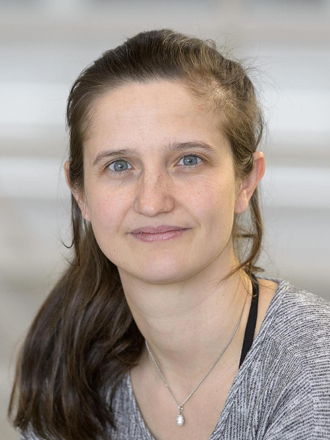
Franziska Lautenschläger received her PhD in Biophysics from the University of Cambridge. During this time she worked mainly on the mechanics of cells, in particular stem cells. She then moved to the Institut Curie, where she became interested in the migration of immune cells. Once independent as a junior professor in Saarbrücken, she combined both interests and worked on the mechanics and resulting migration properties of immune cells. During her time as junior professor, she was also appointed junior group leader at the Leibniz Institute of New Materials, also in Saarbrücken. Since 2020, she has been a full professor of biophysics at Saarland University. Her group tries to understand how the different parts of the cytoskeleton control cellular structure, dynamics and mechanics.

Harnessing Microfluidic Technologies for Advancing Mechanobiology
Affiliation
Lab-on-a-chip Systems Technology Platform, Max-Planck-Insitut für die Physik des Lichts & Max-Planck-Zentrum für Physik und Medizin, Erlangen, Germany
Abstract
Microfluidics is a field that involves the manipulation of small volumes of fluids on the microscale. In recent years, microfluidics has become increasingly relevant due to its ability to provide new insights into cellular and molecular processes, enabling the development of high-throughput and sensitive assays for disease diagnosis, drug discovery and personalized medicine. The flow of cells inside tailored design microfluidic chips can enable a high-throughput and sensitive analysis and sorting of cells based on their physical phenotype. Furthermore, droplet microfluidic technologies provide a unique tool for the development of tailored soft microgel beads mimicking cell physical properties, such as size and elasticity. The development of reliable, simplified and standardized microfluidic-based technologies is essential to enable their broader use and facilitate the translation of research findings into practical applications. Here we illustrate a portfolio of microfluidic chips and standardized cell-mimicking microgel beads rationally designed for enabling the analysis, sorting, and mimics of cells based on their physical properties. Their development was carried out to improve the performance of microfluidic-based technologies by looking at their easy usage, improve performance and reliability. These techniques have the potential to impact various fields, including biophysics and medicine enabling a unique, faster, sensitive, and cost-effective analysis and manipulation of cells.
Biosketch
Salvatore Girardo investigates microfabrication technologies primarily in the area of microfluidics applied to biology and biophysics. He is a Physicist and author on 60 scientific publications and inventor on 3 patents. In 2008 he got a PhD in Nanoscience at the University of Salento (Italy), working on the development of new strategies for the liquid flow control at microscales. In 2013 he joined the Biotechnology Center (TU Dresden, Germany) to setup and manage a new facility, where he implemented novel organ-on-chip platforms, and microfluidic-based methods for cell-mimicking, analysis and treatment. Since 2019 he is the head of the Lab-on-a-chip Systems Technology Platform at the Max Planck Institute for the Science of Light in Erlangen (Germany), working on the development of smart materials and microfluidic technologies mainly applied in the area of mechanobiology.
Marta Urbanska
De novo identification of universal cell mechanics gene signatures
Biotechnology Center, CMCB, TU Dresden, Dresden, Germany; Max Planck Institute for the Science of Light & Max-Planck-Zentrum für Physik und Medizin, Erlangen, Germany; Department of Physiology, Development and Neuroscience, University of Cambridge, Cambridge, UK
Cell mechanical properties determine many physiological functions, such as cell fate specification, migration, or circulation through vasculature. Identifying factors that govern the mechanical properties is therefore a subject of great interest. Here we present a mechanomics approach for establishing links between single-cell mechanical phenotype changes and the genes involved in driving them. We combine mechanical characterization of cells across a variety of mouse and human systems with machine learning-based discriminative network analysis of associated transcriptomic profiles to infer a conserved network module of five genes with putative roles in cell mechanics regulation. We validate in silico that the identified gene markers are universal, trustworthy and specific to the mechanical phenotype, and demonstrate experimentally that a selected target, CAV1, changes the mechanical phenotype of cells accordingly when silenced or overexpressed. Our data-driven approach paves the way towards engineering cell mechanical properties on demand to explore their impact on physiological and pathological cell functions.
Stefan Simm
Explainable artificial intelligence image analysis for blood cell discrimination
Institute of Bioinformatics, University Medicine Greifswald; Institute of Bioanalytics, Coburg University of Applied Science and Art, Greifswald and Coburg, Germany
In terms of decision support processes for high throughput methods it is of major importance to create explainable output for further analysis. In the field of artificial intelligence (AI) and Machine Learning (ML) the addition of explainable components to neural networks (NN) allow the interpretation of classification outputs by finding important features within the input for the decision. As high-throughput imaging approaches of single cells via real time deformability cytometry (RT-DC) create huge imaging datasets we wanted to create an explainable AI (XAI) to discriminate between different cell types within a PBMC blood sample to improve real time sorting without fluorescent labelling of the cell types for later single cell or bulk RNA-Seq analysis on pure cell populations.
For this reason, we used a cohort of ~100 probands with fluorescently labelled blood samples to discriminate between CD19 (B-cells), CD14 (Monocytes), CD3, CD4 or CD8 (T-cells) receptor protein concentrations. After assignment of ~1mio. cells based on an inter-individual 95% quantile threshold we divided in seven cell types and analyzed the possibility to classify them without fluorescence signal information on whether the raw input images via convolutional neutral networks (CNN) or extracted image features like the mechanoprofile via Support Vector Machine (SVM). By the images as well as the mechanoprofile most B cells and Monocytes can be correctly predicted by the CNN and SVM. For the populations of T helper cells (CD3+, CD4+, CD8-) and T suppressor cells (CD3+, CD4-, CD8+), which share a comparable cell size, e-modulus was the most important feature to separate the groups as well as T and B cells differ in the feature: size_y. On the raw images we could observe differences in the importance at the different locations at the membrane to discriminate between the cell types.
