Imaging
Imaging
Tuesday, September 19th, 2023, at 12:00 pm
Chairs: Anja Hauser, Raluca Niesner
Within tissues, cells are constantly exposed to a wide variety of stimuli which impact on their phenotype and function. In addition to “classical” receptor-ligand interactions, these stimuli include a whole range of other cues, for example mechanic and metabolic ones. In this session, we will focus on the analysis of these stimuli, whose importance is increasingly recognized. At the same time, the range of ways to analyze these stimuli continues to expand. The invited speakers in this session will focus on a variety of imaging methods, which enable the quantification of those stimuli in cells and tissues.
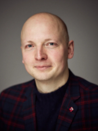
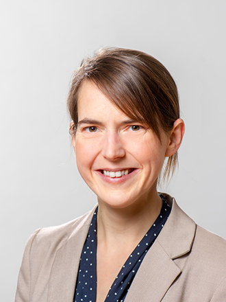
An expert in mechanobiology, Prof. Marco Fritzsche applies sensitive quantitative super-resolution imaging and probing methodologies to analyze the forces occurring during T cell receptor activation at a single cell level, and to understand these processes at a molecular scale. Prof. Nicole Strittmatter´s interest is to combine various imaging technologies in two-and three dimensions. Thus, she focuses on the generation and integration of spatial metabolomics in multi-modal workflows in order to decipher the processes occurring in tissues.

Mechanobiology of T-cell activation in numbers
University of Oxford, Oxford, United Kingdom
Abstract
Mechanobiology is emerging in the biomedical sciences. Recent evidence indicates that immune cells regulate their cell mechanics not only downstream of signalling events triggered by ligand–receptor binding, but that cells dynamically adapt their mechanics in response to external mechanical signals. Quantifying cellular forces has therefore become an contentious challenge across multiple disciplines at the interface of biophysics and immunology. Applying sensitive quantitative super-resolution imaging and force probing methodologies to analyse resting and activated T cells, we demonstrate that the kinetics of the antigen engaging the T-cell receptor controls the nanoscale actin organisation and mechanics of the IS. Using an engineered T-cell system expressing a specific T-cell receptor and stimulated by a range of antigens, force measurements revealed that the peak force experienced by the T- cell receptor during activation was independent of the kinetics of the stimulating antigen. Conversely, quantification of the actin retrograde flow velocity at the IS revealed a striking dependence on the antigen kinetics. Novel ultra-thin superextended lightsheet technology allowed to correlate early calcium activation signalling, IS formation, and effector function. Taken together, these findings suggest that the dynamics of the actin cytoskeleton actively adjusted to normalise the force experienced by the T-cell receptor in antigen specific manner. Consequently, tuning actin dynamics in response to antigen kinetics may thus be a mechanism that allows T cells to adjust the length- and time-scale of T-cell receptor signalling.
Biosketch
Prof Marco Fritzsche is a Associate Professor and Scientific Director of the Oxford-ZEISS Centre of Excellence. He leads the Biophysical Immunology Laboratory between the Rosalind Franklin Institute and the Kennedy Institute for Rheumatology at the University of Oxford, UK. The BPI Laboratory aims to unravel the impact of biophysics and mechanobiology on the human immune response in health and disease. For this mission, the BPI lab develop custom-built microscopy technology at the interface of biophysics and immunology. Prof Fritzsche holds a MSc in theoretical physics and conducted his PhD in experimental biophysics and cell-biology at the London Centre for Nanotechnology at the University College London, UK. He performed his Postdoctoral work at the University of Oxford in close collaboration with the Howard Hughes Medical Institute Janelia Farm, USA.

Spatially resolved metabolomics using mass spectrometry imaging of tissues
Affiliation
School of Natural Sciences – Department of Biosciences, Technische Universität München, Garching/Munich, Germany
Abstract
Small metabolites are the building blocks of life and their sum constitute a chemical fingerprint of the cellular processes at play. Spatial metabolomics using mass spectrometry imaging is performed ex vivo, usually on fresh frozen tissue sections, and enables the localisation of small molecule metabolites, lipids and xenobiotics in two- and three-dimensional context in situ. In multimodal imaging workflows, this additional layer of information can be a crucial piece to generate a holistic view of the processes occurring in complex biological samples such as tissues, enabling the association of metabolites with certain cell types, tissue subregions or phenotypes. We are primarily deploying Desorption Electrospray Ionisation Mass Spectrometry (DESI-MS), an ambient MS technique that is ideally suited for multimodal imaging studies as it requires no sample preparation and leaves the tissue section intact for subsequent imaging-based analysis on the same cell layer. This presentation will showcase the main principles of spatial metabolomics and some applications from our group focussing on tumour metabolism and small molecule drug delivery and efficacy.
Biosketch
Prof. Nicole Strittmatter is a mass spectrometrist with special interest in metabolomics and in situ methods, especially spatial metabolomics using imaging mass spectrometry. Since October 2021, she is an Assistant Professor for Analytical Chemistry at the Technical University of Munich. She studied chemistry at the Justus-Liebig University in Gießen before starting her PhD in Analytical Chemistry at Imperial College London. From 2015-2021, she was working as Imaging Mass Spectrometry Scientist at AstraZeneca, UK, specialising in drug delivery projects.
Laurenz Krimmel
The spatial and unique composition of infiltrating immune cells defines autoimmune- and checkpoint-therapy associated hepatitis
Freiburg University Medical Center, Department of Internal Medicine II, Freiburg, Germany
Background and Aims: Targeting of Immune-checkpoints is commonly used in cancer therapy, but often causes immune-related adverse events. These frequently occur in the form of Immune-checkpoint-blockade associated hepatitis (ICB-Hepatitis).
As the detailed composition and interaction of immune cells in ICB-Hepatitis is unknown and the distinction to autoimmune hepatitis (AIH) remains unclear, our aim was to perform a deep spatial analysis.
Method: We performed a high dimensional, spatially resolved, analysis of immune cell populations in liver biopsies of patients with ICB-Hepatitis (n=15), AIH (n=22) or control tissue (n=10), using a 40-marker Imaging Mass Cytometry (IMC) panel with single-cell resolution (1 μm2). Single cells were segmented utilizing a machine learning pipeline and classified high dimensionally.
Results: Analysis of the immune infiltrate revealed major differences between ICB-Hepatitis and AIH. An unbiased clustering identified disease-specific, phenotypically well defined, immune cell cluster, with unique spatial interactions.
CD8 T cell clustering resulted in enrichment of Ki-67+ Granzyme B+ Clusters in ICB-Hepatitis. AIH, on the contrary, was defined by an exhausted T cell phenotype, as well as tissue resident memory features (Trm). Interestingly, proliferative, and cytotoxic CD8 T cell clusters and the Trm cluster correlated with ALT levels in ICB-Hepatitis and AIH, respectively.
Classification of tissue phenotypes into spatial zones of the liver revealed an enrichment of CD8 T cells around the central vein in ICB-Hepatitis, whereas in AIH a closer interaction with the periportal triad was observed. In ICB-Hepatitis, we observed spatial co-localization of various myeloid clusters and CD8 T cells, especially of proliferative/cytotoxic CD8 clusters and pS6+ macrophages. In AIH, a more prominent involvement of B cells and CD4 T cells became visible.
Conclusion: The composition of the immune infiltrate and its distinct spatial distribution and interaction pattern points towards major differences in the immune mechanisms and pathogenesis driving ICB-Hepatitis and AIH. Our results may have implications for the choice of immunosuppressive approaches in these diseases.
Katja Sallinger
Imaging-based transcriptomics technology identifying predictive biomarkers for relapse in colon cancer stage II
Division of Cell Biology, Histology and Embryology, Gottfried Schatz Research Centre, Medical University of Graz & Center for Biomarker Research in Medicine (CBmed), Graz, Austria
Division of Cell Biology, Histology and Embryology, Gottfried Schatz Research Centre, Medical University of Graz, Graz, Austria and Center for Biomarker Research in Medicine (CBmed), Graz, Austria
Background Therapeutic management of stage II colon cancer remains difficult regarding the decision whether adjuvant chemotherapy should be administered or not. Low rates of recurrence are opposed to chemotherapy induced toxicity and current clinical features are limited in predicting relapse. Predictive biomarkers are urgently needed and therefore we hypothesise that the spatial tissue composition of relapsed and non-relapsed colon cancer stage II patients reveals relevant biomarkers.
Methods The spatial tissue composition of stage II colon cancer patients was examined by a novel spatial transcriptomics technology with sub-cellular resolution, namely in situ sequencing. A panel of 175 genes was designed investigating specific cancer-associated processes such as apoptosis, proliferation, angiogenesis, stemness, oxidative stress, invasion and components of the tumour microenvironment to examine differentially expressed genes in relapsed versus non-relapsed patient samples. Therefore, formalin-fixed paraffin-embedded slides of 5 relapsed and 5 non-relapsed patients were in situ-hybridized and computationally analysed. We identified a tumour gene signature to subclassify tissue into neoplastic and non-neoplastic tissue compartments based on spatial expression patterns generated by in situ sequencing (GTC-tool – Genes-To-Count).
Results The GTC-tool automatically identified tissue compartments that were used to quantify gene expression of biological processes upregulated within the neoplastic vs. non-neoplastic tissue and within relapsed vs. non-relapsed stage II colon patients. Three differentially expressed genes (FGFR2, MMP11 and OTOP2) in the neoplastic tissue compartments of relapsed patients in comparison to non-relapsed patients were identified predicting recurrence in stage II colon cancer.
Conclusions In depth spatial in situ sequencing showed potential to provide a deeper understanding of the underlying mechanisms that are involved in the recurrence of disease and revealed novel potential predictive biomarkers for disease relapse in colon cancer stage II patients. Our developed open-access GTC-tool allowed to accurately capture the tumour compartment and quantified spatial gene expression in colon cancer tissue.
